The Science of Tears
By Daria Zaitseva

Ever cried when watching a movie, chopping onions, or when dirt gets in your eye? The tears rolling down aren’t just saline: They're a sophisticated biological fluid safeguarding your eyes, which contain metabolites, electrolytes, glucose, oxygen, and up to 1,500 proteins, including the most abundant ones associated with anti-inflammatory and antibacterial activity [1, 2]. Tears play a vital role in protecting and lubricating the eye surface. They can even offer insights about your health. Now let’s unpack the science of tears!
What Makes a Tear?
First of all, tears can be classified into three types. “Basal tears” are for housekeeping purposes to keep the eye protected and lubricated all the time. Our eye also secretes “reflex tears” in response to irritants like dust or smoke, and “emotional tears” in response to strong emotions like sadness, anger, and joy [1]. Some scientists suspected that emotional tears could provide an emotional relief based on the discovery that additional proteins and hormones were detected, but current evidence remains inconclusive to this hypothesis [3].
Have you wondered why tears can stay on the eye surface? A thin layer of tear fluid called “tear film” is evenly spread on the eye surface every time we blink. The tear film turns out to be not just a layer of aqueous liquid; it consists of an inner mucin layer, a middle aqueous layer, and an outer lipid layer [1]. The inner mucin layer anchors the middle aqueous layer to the hydrophobic corneal surface. The aqueous layer is crucial for lubricating and protecting the eye surface by flushing away toxins and debris. The outer lipid layer can maintain the thickness of the film by reducing the rate of tear evaporation. Only with these structures can the tear film be firmly attached to the eye surface.
Tear secretion is a carefully controlled process [4]. When sensory afferent nerves of the cornea and conjunctiva detect dryness and irritants [5], they will signal the efferent parasympathetic and sympathetic nerves connected to the lacrimal gland (Figure 1), to induce secretion of electrolytes, water, and proteins to the eye surface [4]. Notably, the sensory input can be modulated by the lacrimal nucleus of the brain, which integrates input from other centers as well, including emotional input, to produce a graded output. A stronger integrated input can induce the secretion of a greater volume of tear by the lacrimal gland. This can explain why tears overflow during emotional episodes, or in response to environmental irritants to flush away deleterious substances. In fact, low levels of nerve stimulation are already enough to produce basal tear to maintain the normal thickness of the tear film.
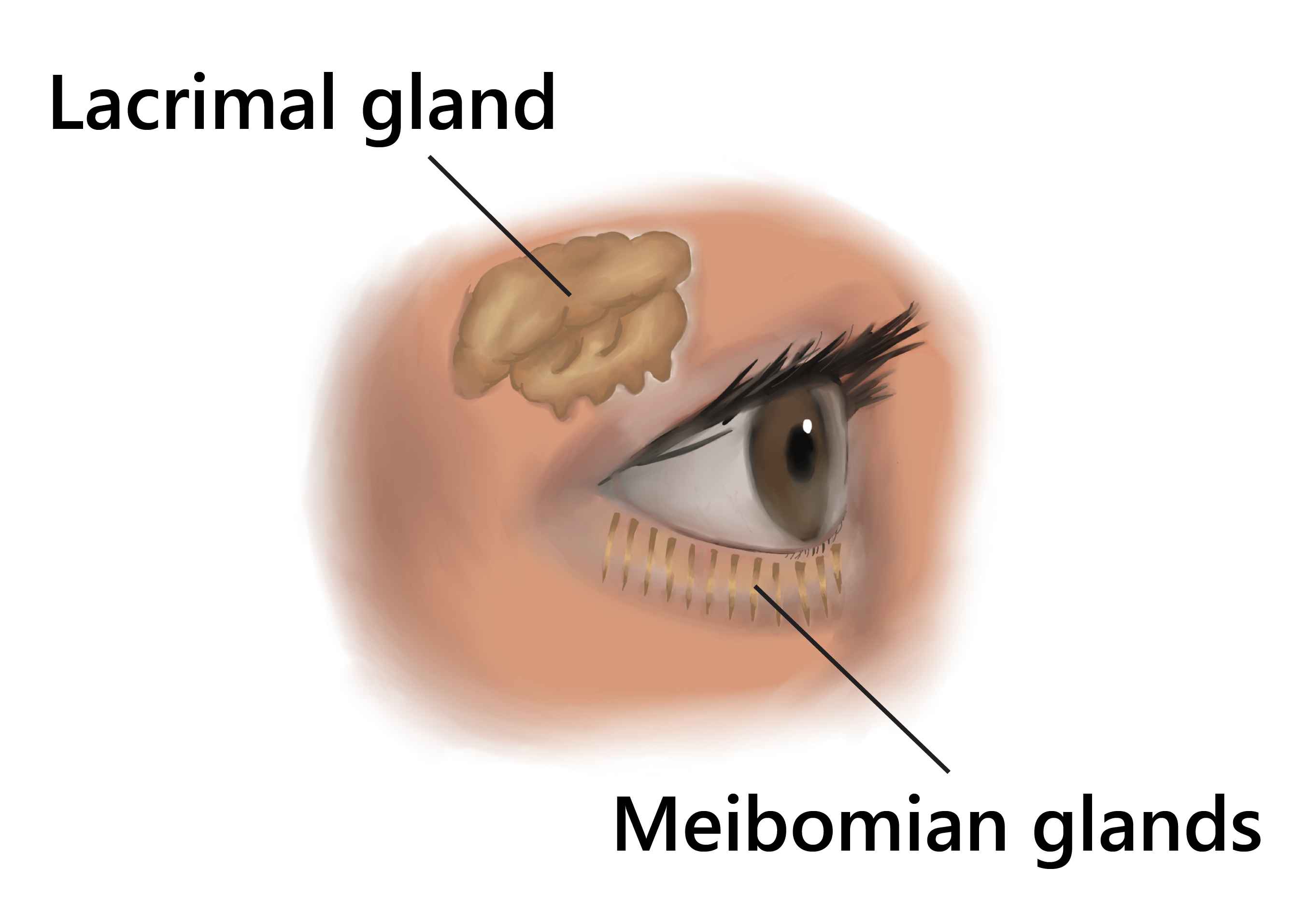
Figure 1 Lacrimal gland and meibomian glands.
Artificial Tears
The uncomfortable sensations of eye dryness can be distressing. Common causes of dry eye include eye strain from prolonged computer use, specific medical conditions, and exposure to smoky or windy environments [6].
To alleviate this discomfort, lubricating eye drops, often referred to as artificial tears, can be beneficial [7]. Most artificial tears consist of aqueous solutions with thickeners such as carboxymethyl cellulose, hyaluronic acid, hydroxypropyl guar, and polyethene glycol to enhance lubrication and prolong their stay on the eye. Natural tears are a non-Newtonian fluid whose viscosity temporarily reduces during each blink to protect the eye surface. Because of the resemblance to natural tears in terms of physical properties, hyaluronic acid is now under extensive research as a promising viscosity-enhancing agent. Other ingredients of artificial tears include electrolytes, pH buffers, antioxidants, and preservatives.
It is also worth noting that such aqueous-based artificial tears work by replenishing the aqueous layer of the tear film. However, lipid-based drops also become increasingly common as they can target the outer lipid layer, relieving dry eye symptoms in individuals whose meibomian gland (Figure 1) cannot properly secrete lipids to maintain the layer [7].
A Cry for Help: Tears in Disease Screening and Health Analysis
As a peripheral body fluid that can be collected in an easy and non-invasive manner, tears have been studied for their potential use in disease screening. By analyzing tear composition, it could be possible to diagnose a disease by quantifying certain biomarkers, in this case biological molecules associated with the disease in question. Scientists are exploring clinical applications for various diseases, from eye diseases like dry eye disease and allergic conjunctivitis, to neurological diseases like Alzheimer’s disease.
For instance, in a subtype of dry eye disease caused by a deficiency in aqueous tear, inflammatory cytokines are synthesized and released to promote inflammation. Multiple studies reported that IL-6, IL-8, and IL-17 are three inflammatory cytokines that could potentially be the biomarkers for the diagnosis of aqueous-deficient dry eye disease [8].
Tear test, if successfully developed, could also help with the diagnosis of another eye disease, allergic conjunctivitis (AC). Type IV AC is associated with prolonged exposure to allergens, but it is often mistaken for seasonal type I AC in clinical practice. A quick test to quantify the amount of IgE in tear fluid could reliably differentiate the two conditions because low IgE levels are found to be indicative of type IV AC. The test will enable physicians to administer appropriate medication to the patients [9].
Non-invasive tear tests could also become an easy screening method for various neurological diseases because the elevated level of biomarkers in cerebrospinal fluid is also observed in tears in some cases. For example, TNF-alpha and alpha 1-antichymotrypsin are two such biomarkers for Parkinson’s disease and multiple sclerosis, respectively [10]. Scientists are also making efforts to identify reliable biomarkers for Alzheimer’s disease. If tear-based screening methods can be developed and commercialized eventually, we will be able to promote population screening in the community. Early diagnosis and treatment can improve the quality of life for both the patients and their caregivers [11].
As for tear-based biodevices, a recent study suggested the possibility for diabetic patients to continuously monitor their tear glucose level with a smart contact lens [12]. The previous challenge of using tear glucose level as an alternative indicator for blood glucose was that single measurement using conventional tear collection methods, such as filter paper strip and capillary tube, always undesirably induce the generation of reflex tears, which will interfere with the results. By embedding an antenna, a glucose sensor and an NFC chip in the soft contact lens, the research team could continuously monitor the glucose level in basal tears, with the ability to transfer real-time data to a mobile device.
While tears may contain a wide array of biomarkers that can reveal our health status, there is still a long way to go before relevant technologies can reach the clinic. With extensive research efforts working on the identification of biomarkers and the development of smarter biodevices, tears can one day become a powerful indicator of our health.
The Shape of Tears One way to artistically study tears is to observe them under a microscope – by observing the air-dried salt crystals or the tear fluid compressed between a microscopic slide and a coverslip [13]. A photographer, Rose-Lynn Fisher, created a project called “The Topography of Tears,” in which she captured the diverse morphology of tears shed by herself and her friends on various occasions. More about the project: https://rose-lynnfisher.com/tears.html |
References
[1] Chang, A. Y., & Purt, B. (2023, June 5). Biochemistry, Tear Film. StatPearls. StatPearls Publishing.https://www.ncbi.nlm.nih.gov/books/NBK572136/
[2] Vera-Montecinos, A., Pardo, C. C., Hernández, M., Saldivia, P., Nourdin, G., Elizondo-Vega, R., Sánchez, E., Amulef, S., Koch, E., Vargas, C., & Oyarce, K. (2025). High throughput tear proteomics with data independent acquisition enables biomarker discovery in allergic conditions. Scientific reports, 15(1), 31181. https://doi.org/10.1038/s41598-025-17105-y
[3] Collier, L. (2014, February). Why we cry. Monitor on Psychology, 45(2), 47. https://www.apa.org/monitor/2014/02/cry
[4] Dartt, D. A. (2009). Neural regulation of lacrimal gland secretory processes: Relevance in dry eye diseases. Progress in Retinal and Eye Research, 28(3), 155–177. https://doi.org/10.1016/j.preteyeres.2009.04.003
[5] Meng, I. D., & Kurose, M. (2013). The role of corneal afferent neurons in regulating tears under normal and dry eye conditions. Experimental Eye Research, 117, 79–87. https://doi.org/10.1016/j.exer.2013.08.011
[6] Pagan-Duran, B. (2022, February 9). Lubricating Eye Drops for Dry Eyes. EyeSmart, American Academy of Ophthalmology. https://www.aao.org/eye-health/treatments/lubricating-eye-drops
[7] Semp, D. A., Beeson, D., Sheppard, A. L., Dutta, D., & Wolffsohn, J. S. (2023). Artificial Tears: A Systematic Review. Clinical Optometry, 15, 9–27. https://doi.org/10.2147/OPTO.S350185
[8] Fong, P. Y., Shih, K. C., Lam, P. Y., Chan, T. C. Y., Jhanji, V., & Tong, L. (2019). Role of tear film biomarkers in the diagnosis and management of dry eye disease. Taiwan Journal of Ophthalmology, 9(3), 150–159. https://doi.org/10.4103/tjo.tjo_56_19
[9] Shang, X., Zhang, Y., Luo, S., Liu, M., Li, H., Fang, X., Xie, Z., Xiao, X., Yang, Z., Lin, Y., & Wu, H. (2025). Tear IgE point-of-care testing for differentiating type I and type IV allergic conjunctivitis. Frontiers in Medicine, 12. https://doi.org/10.3389/fmed.2025.1577656
[10] Gijs, M., Ramakers, I. H. G. B., Visser, P. J., Verhey, F. R. J., van de Waarenburg, M. P. H., Schalkwijk, C. G., Nuijts, R. M. M. A., & Webers, C. A. B. (2021). Association of tear fluid amyloid and tau levels with disease severity and neurodegeneration. Scientific Reports, 11(1), 22675. https://doi.org/10.1038/s41598-021-01993-x
[11] Kalló, G., Emri, M., Varga, Z., Ujhelyi, B., Tőzsér, J., Csutak, A., & Csősz, É. (2016). Changes in the Chemical Barrier Composition of Tears in Alzheimer's Disease Reveal Potential Tear Diagnostic Biomarkers. PLoS one, 11(6), e0158000. https://doi.org/10.1371/journal.pone.0158000
[12] Park, W., Seo, H., Kim, J., Hong, Y., Song, H., Joo, B. J., Kim, S., Kim, E., Yae, C., Kim, J., Jin, J., Kim, J., Lee, Y., Kim, J., Kim, H. K., & Park, J. (2024). In-depth correlation analysis between tear glucose and blood glucose using a wireless smart contact lens. Nature Communications, 15(1), 2828. https://doi.org/10.1038/s41467-024-47123-9
[13] Fisher, R. (n.d.). The Topography of Tears. https://rose-lynnfisher.com/tears.html